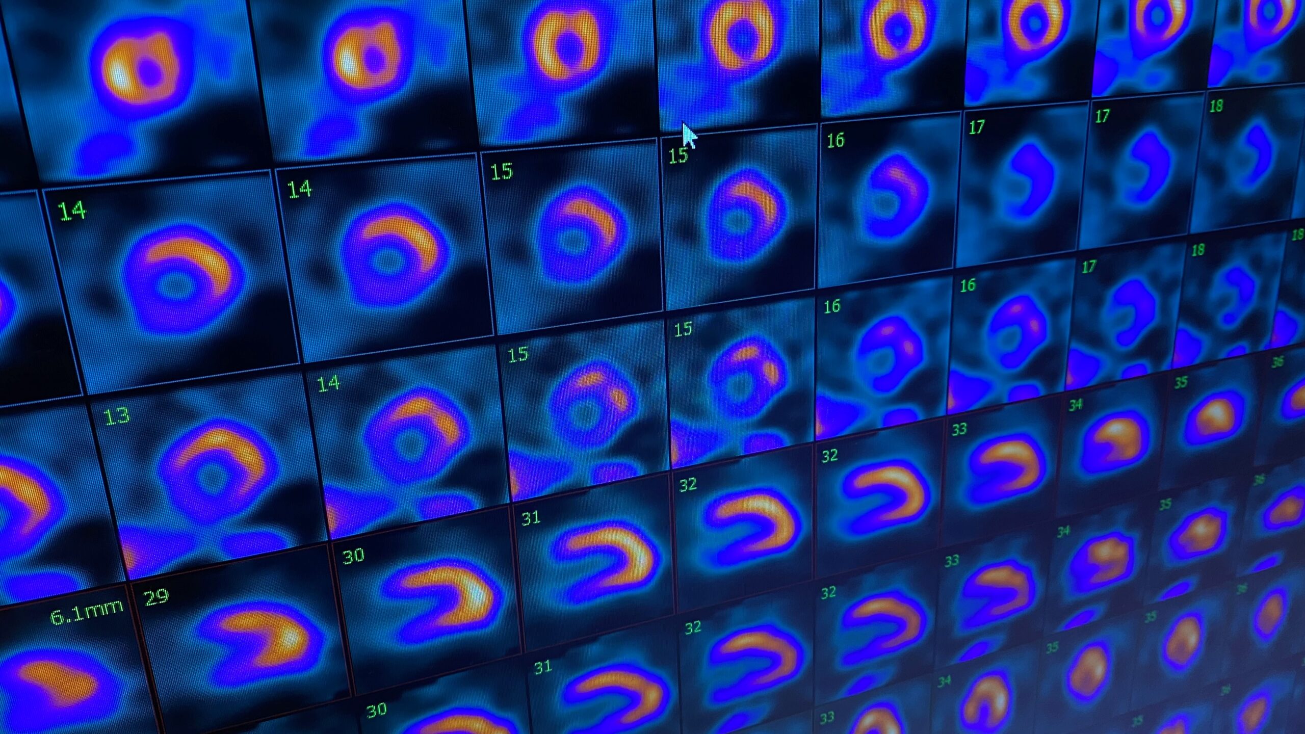
Cardiac PET/CT
A cardiac positron emission tomography (PET) scan is a noninvasive, nuclear medicine diagnostic test that allows your doctor to see how blood flows to your heart muscle, as an indicator of how well your heart and its tissues are functioning. Cardiac PET scans are useful for diagnosing conditions like a blockage in the arteries that supply blood to the heart (the coronary arteries) and for determining how much damage was caused by a heart attack —even one you might not know you’ve had.
Cardiac computed tomography (CT, or “cat scan”) imaging, another noninvasive diagnostic test, takes many x-ray pictures of thin slices of your heart and then uses a computer to merge these images together.
Scanners that combine PET and CT produce structural images of much higher quality and have advanced software for measuring how much blood is flowing to each area of your heart. Compared with traditional nuclear stress tests, hybrid PET/CT imaging is much more accurate, which helps your doctor diagnose your condition more precisely and make better treatment decisions. A cardiac PET/CT scan may also be enough for your doctor to determine whether coronary artery disease is present or absent, thus avoiding further invasive testing.
The imaging scan is safe and typically takes less than 45 minutes in the outpatient setting.
PET/CT at The Texas Heart Institute Center for Cardiovascular Care
At The Texas Heart Institute Center for Cardiovascular Care, our constant goal is to provide the highest-quality care for our patients. Our state-of-the-art PET/CT scanner allows us to do just that: Its advanced diagnostic capabilities, speed, power, and flexibility in cardiac imaging generate highly accurate anatomical reconstructions, giving us the tools we need to investigate cardiac conditions. At our new patient-focused cardiovascular center, we provide exceptional care to patients and visitors from the Houston area and around the world.
The PET/CT scanner and diagnostic suite at the Center for Cardiovascular Care provides the most advanced imaging available today. As one of the few centers offering Heartsee™ technology, we can obtain highly accurate data to help detect flow abnormalities. The imaging suite also includes five echocardiogram rooms to perform cardiac ultrasonography; two peripheral vascular imaging rooms to assess arterial and venous disease; four cardiac stress-test rooms; a SPECT (single-photon emission computed tomography) scanner; and a general radiology room.
What should I do before having a PET/CT scan?
Inform your doctor if you are pregnant or nursing, because the radiation could be harmful to your baby or nursing infant. Alternative imaging exams are available.
Talk to your doctor ahead of time about all the medications you are taking. Bring a list of them with you. You should especially avoid taking any theophylline-containing medications for at least 48 hours before your scan.
You must fast before the test. For 6 hours before the test, you must not eat anything. Drink plenty of water up to 2 hours before the test.
Do not drink any caffeinated beverages for 24 hours (ideally, 48 hours) before the test.
If you have diabetes, talk to your doctor about food and insulin needs, because fasting before your scan can affect your blood sugar levels.
Even though the PET/CT scanner is more open than traditional MRI scanners, do tell your doctor if you have a fear of enclosed spaces (claustrophobia). The doctor may give you medicine before the test that will help you relax.
Wear loose, comfortable clothing. Do not wear a dress, clothing with metal buttons or metal snaps, or jewelry.
What happens during my PET/CT scan?
Heart monitor leads will be taped to your chest.
For the PET portion of the test, a small amount of radiotracer (radioactive material) will be injected into your arm. The radiotracer deposits itself in your organs and tissues, which allows the scanner to detect the energy it emits and produce 3D images. The amount of radiation is minimal. It breaks down quickly in your body.
You may be injected with an agent to simulate stress conditions in your heart. This medication is safe.
Once the radiotracer has had time to move throughout your body, you will lie down on an exam table that will move through a large tunnel (the PET/CT scanner). You must lie still so that the images will be clear. The scanner will automatically adjust for breathing and heart motion.
The combined PET/CT image will show what is happening in the cells (from the PET scan) and where it is happening (from the CT scan).
Your doctor will share the results with you when they become available, which may take 24-72 hours.
After the test is over, you may eat, drink, and go back to your normal activities right away.
